Conducting polymers (CPs) are an exciting group of organic biomaterials which change their properties upon application of a suitable stimulus. These polymers are synthesized by either chemical, electrochemical or vapour phase polymerization (VPP). VPP has been reported to produce highly organized biocompatible CP structures with high conductivities [1]. CPs are redox responsive materials; high surface area structures facilitate interactions between the CP and the surrounding environment eliciting faster and larger magnitude responses. In this study, a novel approach was taken to fabricate highly porous poly(3,4-ethylenedioxythiophene) (PEDOT) structures over a sacrificial template using VPP. PEDOT is a robust CP reportedly resistant towards over-oxidation thus an ideal candidate for long-term use in biological conditions [2]. It has been investigated for various biomedical applications including biosensing and stimuli-responsive drug delivery [2][3][4].
A sacrificial polystyrene (PS) colloidal template was first deposited on a borosilicate glass substrate by vertical deposition. Following deposition these colloids were heat cured for 1 hr at 90 °C, and then an oxidant solution of iron tosylate was spun coated at 1500 rpm for 25 sec. VPP was conducted in a vacuum oven at 45 mbar and 35 °C for 1 hr while the EDOT reservoir was heated to 45 °C. Structures were characterized by x-ray photoelectron spectroscopy (XPS), atomic force microscopy (AFM), scanning electron microscopy (SEM), fourier transformed infra-red spectroscopy (FTIR), cyclic voltammetry (CV) and electrochemical impedance spectroscopy (EIS). Extracts collected from phosphate buffer saline (PBS) following 100 redox cycles between -0.9 V and 1.4 V at 100 mV/sec were analysed for cytotoxicity using human retinal pigment epithelial (ARPE-19) cells.
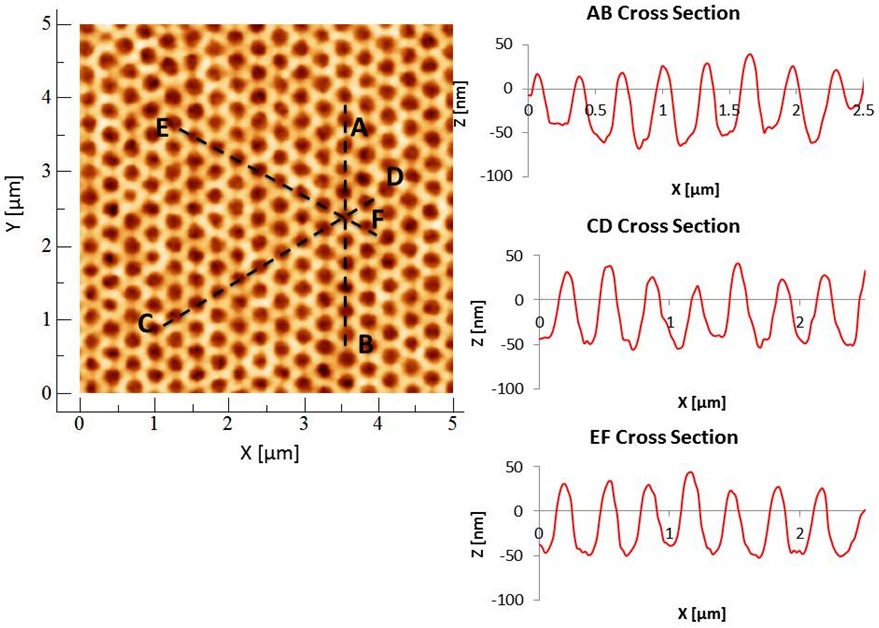
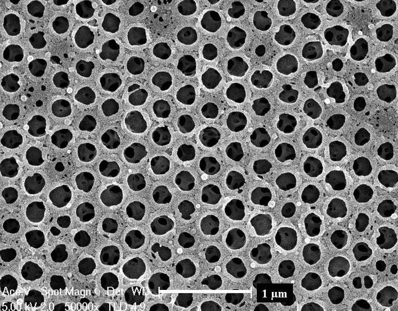
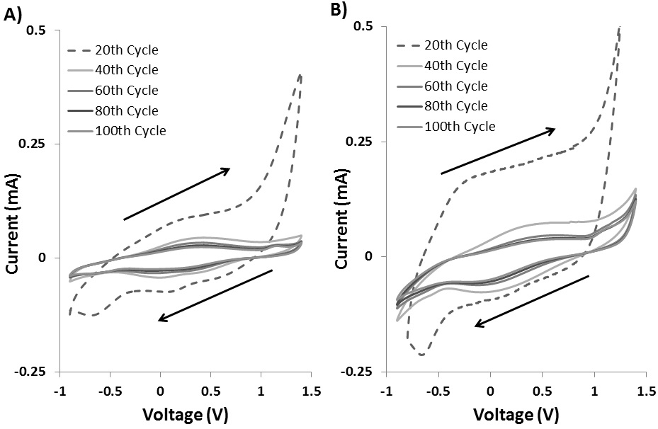
AFM and SEM revealed the highly porous honey comb nature of these structures [Fig. 1, Fig. 2]. XPS and FTIR data showed the composition of these materials related well with that of PEDOT. These materials retain their electroactive properties through many cycles of reduction and oxidation as demonstrated by CV [Fig. 3]. EIS showed that these novel structures offer 75 ohm impedance at 100 KHz (common frequency for wireless transcutaneous electricity transmission) when compared to 776 ohm at the same frequency for non-porous VPP PEDOT films for the same area and polymerization time. Cell viability studies were able to demonstrate that extracts from these structures had minimal toxic effects on the human cells showing cell viability of about 90% when compared to PBS.
This is the first report of the successful fabrication of porous PEDOT structures prepared through VPP over a sacrificial colloidal template. The produced structures have a high surface area, low impedance and minimal cytotoxicity. These exciting structures offer reproducible redox ability over several cycles desirable for long-term use in biomedical devices for biosensing and drug delivery.
References:
[1] E.M. Stewart, M. Fabretto, M. Mueller, P.J. Molino, H.J. Griesser, R.D. Short, G.G. Wallace, Cell attachment and proliferation on high conductivity PEDOT-glycol composites produced by vapour phase polymerisation, Biomater Sci-Uk 1 (2013) 368-378.
[2] H. Yamato, M. Ohwa, W. Wernet, Stability of Polypyrrole and Poly(3,4-Ethylenedioxythiophene) for Biosensor Application, J Electroanal Chem 397 (1995) 163-170.
[3] S. Venkatraman, J. Hendricks, Z.A. King, A.J. Sereno, S. Richardson-Burns, D. Martin, J.M. Carmena, In Vitro and In Vivo Evaluation of PEDOT Microelectrodes for Neural Stimulation and Recording, IEEE Trans. Neural Syst. Rehabil. Eng. 19 (2011) 307-316.
[4] J.A. Chikar, J.L. Hendricks, S.M. Richardson-Burns, Y. Raphael, B.E. Pfingst, D.C. Martin, The use of a dual PEDOT and RGD-functionalized alginate hydrogel coating to provide sustained drug delivery and improved cochlear implant function, Biomaterials 33 (2012) 1982-1990.